

Achieving successful treatment outcomes relies upon using the best diagnostic technology to develop the most appropriate plans of care. With the advanced and versatile imaging capabilities offered by the i-CAT system, it’s not only possible to obtain detailed 3D views of a patient's teeth, jaws, and the surrounding anatomy but also conventional 2D panoramic views when needed. As a "best in class" high-resolution CBCT scanner (cone-beam computed tomography), the i-CAT system provides the most reliable and accurate images to facilitate precise and effective care.
An exceptionally designed and engineered system, i-CAT imaging system offers minimal-exposure imaging options and can very comfortably obtain distortion-free, detailed views from several angles. The i-CAT system provides essential information for multiple types of analyses, as well as the assessment of maxillofacial disorders or pathology. It is also most useful in surgical planning, including the accurate placement of dental implants.
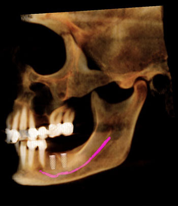
3-Dimensional software supports comprehensive case planning and the precise visualization of all anticipated treatment changes and improvements. It enhances patient-doctor communication by enabling patients to preview their treatment as well as the outcome so that they can feel fully informed of all their options in care.
For the oral and maxillofacial surgeon, 3-dimensional software optimizes treatment planning by integrating information from multiple sources and allows the visualization of tissue changes in all three planes of space. This software not only animates the patient’s skeletal and facial changes in real-time, but it also permits accurate virtual model surgery and outputs information for precise surgical guides. Compatible with other platforms, 3-dimensional software also supports the construction of custom-fitted appliances and restorations.
Digital radiography utilizes computer technology and digital sensors for the acquisition, viewing, storage, and sharing of radiographic images. It offers several advantages over the older traditional film based methods of taking x-rays. The most significant of theses advantages is that digital radiography reduces a patient’s exposure to radiation. Other benefits are that images can be viewed instantly after being taken, can be seen simultaneously as needed by multiple practitioners, and can be easily shared with other offices. Digital x-rays are also safer for the environment as they do not require any chemicals or paper to develop.
An electronic pad, known as a sensor is used instead of film to acquire a digital image. After the image is taken, it goes directly into the patient’s file on the computer. Once it is stored on the computer, it can be easily viewed on a screen, shared, or printed out.
The digital equivalent of a paper patient chart, electronic medical records (EMR) allow for seamless record keeping and facilitate the sharing of information between healthcare professionals and other providers for more efficient care.
With an electronic medical record, a patient’s medical history and relevant medical data are instantly available to the treating provider. This feature also boosts the quality and safety of patient care as information on previous prescriptions of medications or allergic reactions can be immediately viewed. We can also provide patients with clinical summaries, including information on the diagnosis, care provided, medications prescribed, upcoming/follow-up appointments and related medical advice.

Digital optical impressions significantly increase efficiency, productivity and accuracy, and make it possible for us to e-mail the virtual impression to your dentist and or laboratory, rather than send a traditional impression or stone model via regular mail. Also, digital impressions can be used to make surgical guides for implant placement, thereby making the implant process efficient and accurate.
Other benefits of digital impressions include:
